過敏性濕疹病人體內金黃色葡萄球菌內毒素對組胺和白三烯釋放的研究
過敏性濕疹病人體內金黃色葡萄球菌內毒素對組胺和白三烯釋放的研究
(國外學術刊物發(fā)表)
Staphylococcus aureus Enterotoxins Induce Histamine and Leukotriene Release in Patients with Atopic Eczema
K. Neuber, J. Wehner, K. Hakansson, and J. Ring
Abstract
Peripheral blood basophils from patients with atopic eczema stimulated with the staphylococcal enterotoxins (SE) A, B, D, E and toxic shock syndrome toxin 1 (TSST-1) secreted significantly higher amounts of histamine and leukotriene C4 (LTC4 ) than healthy controls. Furthermore, priming experiments with IL-3, IL-8 and GM -CSF followed by stimulation with enterotoxins showed additional histamine and LTC4 release in the group of atopic eczema patients. Histamine and leukotriene generation from atopic basophils stimulated with staphylococcal enterotoxins may indicate a role of these toxins as possible allergens in at least a subgroup of patients with atopic eczema.
Introduction
Normal as well as diseased skin of patients with atopic eczema (AE) is severely colonized with Staphylococcus aureus [1]. Recently, it has been shown that the majority of S. aureus strains isolated from the skin of patients with AE produce staphylococcal enterotoxins (SEA, B, C, D, E) or toxic shock syndrome toxin-1 (TSST-1). About 50% of the patients have specific IgE directed to staphylococcal toxins [2]. The activation of T-cells by staphylococcal superantigens results in the release of a TH2-like cytokine pattern (IL-4, IL-5) in vitro [3] which, via induction of several effector cells, may be the cause of increased IgE-synthesis and eosinophilia. In this study the influence of staphylococcal enterotoxins (SEs) on histamine and leukotriene release of basophils was studied.
Material and Methods
Patients and Controls. Basophils were obtained from healthy volunteers (n = 9) and from patients with AE (n = 17). The AE diagnosis was performed according to the criteria of Hanifin and Rajka [4]. All patients had a skin colonization with S. aureus as was determined by skin smears. The donors did not receive systemic steroid or antihistamine treatment.
Blood Collection and Cell Preparation. Peripheral blood leukocytes (PBL) were obtained by dextrane-sedimentation for 60-90 min. After three washing steps, the pellet was resuspended in 5 ml HEPES-ACM buffer (pH 7.4). Stimulation Conditions. As stimuli served S. aureus enterotoxins (SEA, SEB, SED, SEE and TSST-1) obtained from Toxin Technology (Sarasota, USA) diluted in carbonate buffer (pH 8.0). For priming experiments interleukins-3, 8 and granulocyte-macrophage colony-stimulating factor (GM -CSF), all obtained from Biermann DPC (Bad Nauheim, Germany), were used. As controls served spontaneous as well as anti-IgE (10-2 M) induced histamine and leukotriene release. Total leukocyte histamine content was determined by sonication of cells for 10 s. Aliquots (500 μl) of leukocyte suspension (1.0-1.5 x10⁷/ml) were stimulated with SEs (100 ng/500 μl) for 30 min at 37 °C in duplicates. For priming experiments leukocytes were prestimulated (10 min) with interleukins in the following concentrations: IL-3 (500 ng/500 μl), IL-8 (40 ng/500 μl) and GM-CSF (20ng / 500 μl). Afterwards, toxins were added for 30 min at 37 °C. After incubation period reactions were stopped by adding 500 μl 0.01 N TFA (C2HF3O2).Supernatants were stored at –20 °C.
Histamine and LTC4 ELISA. Histamine and LTC4 contents of supernatants were determined by ELISA obtained from IBL (Hamburg, Germany) according to the prescription of the manufacturer. The release of histamine, either spontaneously or as induced by stimuli, was recorded as a percentage of the total net leukocyte histamine release.
Analysis of Data. All calculations were performed using the SPSS Statistics package (SPSS, Vers. 5.02). Results were analysed using the nonparametric Wilcoxon signedranks test for paired data or the Wilcoxon non-paired rank-sum test for nonpaired data. Only p-values below 0.05 were accepted as significant.
Results
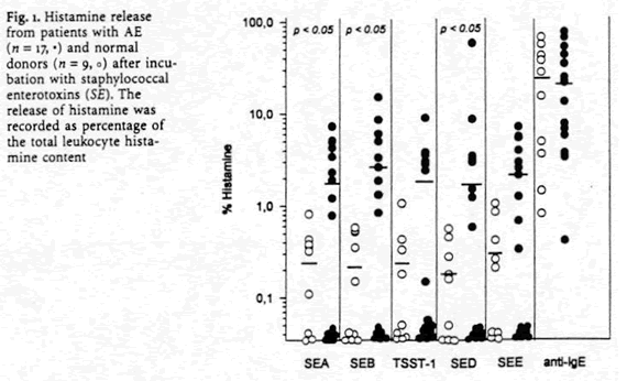
Enterotoxin Induced Histamine and LTC4 Release. Basophils from healthy subjects did not release more than 2 % histamine to any staphylococcal toxin. In contrast, 14 AE patients (82 %) showed relevant histamine release upon stimulation with one or more staphylococcal enterotoxins (Fig. 1).

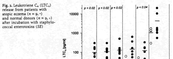
Leukocytes from patients with AE generated significantly higher amounts of LTC4 (Fig. 2) after stimulation with SEA (p<0.02), SEB (p<0.02), TSST-1 (p<0.02) and SEE (p<0.04).

Histamine and LTC4 Release After Priming with IL-3, IL-8 and GM-CSF. Priming of atopic basophils with IL-3, IL-8 or GM -CSF induced a significantly enhanced histamine release compared to stimulation with enterotoxins alone (Table 1). In normal donors no histamine release over 2 % was observed after cytokine priming and incubation with enterotoxins. In healthy volunteers priming with interleukins (3, 8) and GM -CSF did not enhance enterotoxin induced LTC4 generation. However, in patients with AE priming of basophils led to an increase of LTC4 secretion by 875 % for IL-3, 187 % for IL-8 and 412 % for GM -CSF compared to cells stimulated without interleukins (Table 1).

Discussion
These data clearly show that SE stimulated basophils from patients with AE secrete significantly higher amounts of histamine and LTC4 than healthy donors. Furthermore, priming of basophils with IL-3, IL-8 and GM-CSF increased mediator release in AE. Recently, an increasing number of data support the suggestion that parts of the
cutaneous microflora (e. g.) Staphylococcus aureus, Pityrosporum ovale) acting as permanent stimuli for allergic skin reactions can be an important trigger for the exacerbation of AE [3,5]. SEs have been demonstrated to be stimuli of TH2 cells in AE inducing IL-4 and IL-5 secretion and IgE synthesis [3].
Histamine and LTC4 are very important mediators of inflammation in allergic diseases [6]. Therefore, induction of mediator release by SEs in AE supports the assumption that these toxins may trigger allergic inflammation. Since AE is associated with superficial S. aureus skin colonization, secreted enterotoxins would be expected to penetrate the skin. Binding of SEs to cell surface bound specific IgE antibodies can mediate histamine release from basophils and mast cells. This may lead to local release of mediators and cytokines resulting in late-phase reactions and itch. In control experiments with buffer not containing Ca2+ SEs as well as cytokines failed to induce histamine secretion even in patients with AE (data not shown). These results are similar IgE mediated histamine and LTC4 formation.
One of the most important modulating effects of cytokines on inflammatory cells is their capacity to prepare the cells for more efficient mediator release, a phenomenon called “priming”. Blood basophils markedly increase their capacity to release preformed histamine or to form de novo LTC4 following short incubation in vitro with IL-3, IL-8 and GM-CSF [7]. The fact that SE induced mediator release in AE is significantly increased by cytokines supports the assumption that hypersensitivity reaction to these toxins are pathophysiologically relevant in at least a subgroup of patients with AE. Acknowledgements. The authors wish to thank Ingrid Köhler and Johanna Grosch for excellent technical assistance.
References
1. Ring J, Abeck D, Neuber K. Atopic eczema: role of microorganisms on the skin surface. Allergy 1992; 47:265-269
2. Leung DYM, Harbeck R, Bina P, Reiser RF, Yang E, Yang DA, Norris DA, Hanifin JK, Sampson HA. Presence of IgE antibodies to staphylococcal exotoxins on the skin of patients with atopic dermatitis. J Clin Invest 1993; 92:1374-1380
3. Neuber K, Steinrücke K, Ring J. Staphylococcal enterotoxin B affects in vitro IgE synthesis, interferon-g, interleukin-4 and interleukin-5 production in atopic eczema. Int Arch Allergy Immunol 1995; 107:179-182
4. Hanifin J, Rajka G. Diagnostic features of atopic dermatitis. Acta Derm Venereol (Stockh) 1980; 92:44-47
5. Kröger S, Neuber K, Gruseck E, Ring J, Abeck D. Pityrosporum ovale extracts increase interleukin-4, interleukin-10 and IgE synthesis in patients with atopic eczema. Acta Derm Venereol (Stockh) 1995; 75:357-360
6. Ruzicka T, Ring J. Role of inflammatory mediators in atopic eczema. In: Handbook of atopic eczema. Eds.: Ruzicka T, Ring J, Przybilla B. Berlin: Springer, 1991; p 245-255
7. de Weck AL, Dahinden CA, Bischoff S. Cytokines in the regulation of allergic inflammation. In: Advances in allergology and clinical immunology: the proceedings of the XVth congress of the European Academy of Allergology and Clinical Immunology. Eds.: Godard P, Bousquet J, Michel FB. Park Ridge: The Parthenon Publishing Group. 1992; p 67-74
Copyright(C) 1998-2024 生物器材網 電話:021-64166852;13621656896 E-mail:[email protected]


