Primary cultures of intrahepatic bile duct epithelial cells isolated and cultured
1. Liver was surgically removed and perfused via the hepatic vein.
2. The liver was perfused with 0.05% (wt/vol) collagenase in primary cell system after flushing with Hanks balanced salt solution, pH 7.4, containing 50 mM EGTA.
3. The collagenase-digested liver was then minced into small fragments and allowed to incubate in primary cell system containing 0.05% (wt/vol) collagenase and 0.05% (wt/vol) hyaluronidase for 30 min at 37°C.
4. The minced tissue was washed several times in primary cell system and passed through a crude sieve to obtain a total liver cell suspension.
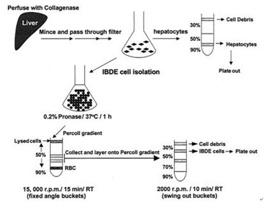
5. An aliquot of the cell suspension was processed for primary hepatocyte culture while the rest of the hepatocytes in the total liver cells were lysed by incubation in 100 ml of 0.2% (wt/vol) pronase in primary cell system for 45 min at 37°C.
6. DNase I was then added to a final concentration of 100 μg/ml to digest released cellular DNA, which may cause cell clumping.
7. After incubation for a further 15 min, the enzymes were removed from the cell suspension by several washes in primary cell system.
8. The cells were then resuspended in a minimal volume of medium and layered onto a Percoll gradient comprised of 20 ml of 50% (vol/vol) and 5 ml of 90% (vol/vol) isotonic Percoll.
9. The gradients were centrifuged at 15,000 rpm for 15 min at room temperature (RT) in a centrifuge to separate lysed hepatocytes and other cell debris from the nonparenchymal cells.
10. Cells banding lower down the 50% Percoll gradient were collected, washed, and overlaid onto a second gradient comprised of 2.5 ml of 30% (vol/vol), 2.5 ml of 50% (vol/vol), 2.5 ml of 70% (vol/vol), and 1 ml of 90% (vol/vol) isotonic Percoll; Percoll density marker beads were also layered on top of a parallel gradient.
11. The gradient was centrifuged at 2,000 × g for 15 min at RT, and cells banding at each Percoll interface were collected, washed, and resuspended in growth medium, which was primary cell system containing 5% (vol/vol) fetal calf serum, 100 μg of soybean trypsin inhibitor/ml, 0.02% (wt/vol) glucose, 450 ng of hydrocortisone 21-hemisuccinate/ml, 1× insulin-transferrin-selenium (ITS)-A supplement, 100 U of penicillin/ml, 100 μg of streptomycin/ml, 1.5% (vol/vol) dimethyl sulfoxide, and 1.5% (vol/vol) duckling sera.
12. Cells found banding at each Percoll interface were designated from the top to the bottom of the gradient as fractions F1 (1.04/1.06 g/cm3), F2 (1.06/1.08 g/cm3), and F3 (1.08/1.11 g/cm3).
13. The cells from each interface were seeded onto 12-well plates and coverslips (12-mm diameter) that had been thinly coated with rat tail collagen according to the manufacturer's instructions.
14. The cells were maintained by medium changing every second day.
15. For primary cultures of duck hepatocytes, an aliquot of total liver cells from the above-mentioned IBDE cell isolation procedure was overlaid onto a Percoll gradient comprised of 30% (vol/vol), 50% (vol/vol), and 90% (vol/vol) isotonic Percoll.
16. The gradient was centrifuged at 2,000 × g for 10 min at RT.
17. A yellow layer of cells banding at the 50% and 90% Percoll interface was collected, washed several times in growth medium, and seeded onto 12-well plates and coverslips (12-mm diameter). The culture medium was changed every second day.
Schematic diagram of the method used for the isolation of IBDE cells from duckling liver.

References
1. Jia-Yee Lee, Janetta G. Culvenor, Peter Angus, Richard Smallwood, Amanda Nicoll, and Stephen Locarnini. Duck hepatitis B virus replication in primary bile duct epithelial cells. J Virol. 2001; 75: 7651–7661.
2. Wang Y, Luscombe C, Bowden S, Shaw T, Locarnini S. Inhibition of duck hepatitis B virus DNA replication by antiviral chemotherapy with ganciclovir-nalidixic acid. Antimicrob Agents Chemother. 1995; 39: 556–558.
3. Sirica AE, Gainey TW. A new rat bile ductular epithelial cell culture model characterized by the appearance of polarized bile ducts in vitro. Hepatology. 1997; 26: 537–549.


